Edible Cultivated Meat
Yuya Morimoto, Mai Furuhashi, Kaori Aibe, Byeongwook Jo, Shoji Takeuchi
Food and Bioprocess Technology, vol. 19, 416, 2026
View Research Details
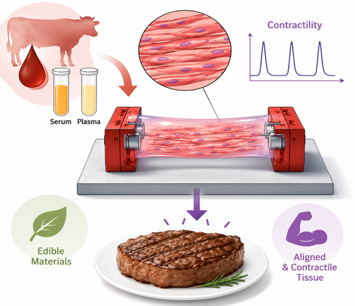
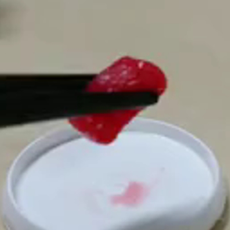
We developed a method for producing edible cultivated steak using only edible bovine blood-derived components: serum and plasma. Plasma was used as the scaffold material, while serum was used as a culture-medium component, eliminating dependence on conventional non-edible materials such as collagen. Using a modular assembly approach in which individually prepared muscle tissues were stacked together, we constructed a large tissue approximately 10 cm in length. Electrical stimulation experiments confirmed that the tissue was mature, with aligned muscle fibers and contractile properties resembling those of living muscle. In a tasting evaluation, the tissue was considered similar to conventional meat in elasticity and chewiness, although further improvements are needed in flavor and juiciness. This work represents an important step toward the commercialization of clean and ethically produced cultivated meat.
Cultivated Steak
Formation of contractile 3D bovine muscle tissue for construction of millimetre-thick cultured steak
(*equal contribution) Mai Furuhashi*, Yuya Morimoto*, Ai Shima, Futoshi Nakamura, Hiroshi Ishikawa, Shoji Takeuchi
npj Science of Food, vol. 5, 6, 2021
View Research Details
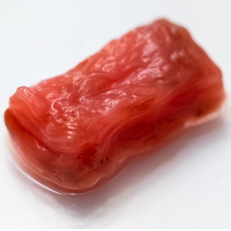
To help address potential future meat shortages, we developed a method for producing millimetre-thick cultivated steak with highly aligned muscle fibers. Hydrogels containing bovine cells were formed into modules with a unique striped structure. These modules were then stacked using a modular assembly approach, enabling the construction of larger tissues. This method promoted strong unidirectional alignment of the muscle fibers while enhancing their maturation and contractile function. Experimental evaluation showed that the resulting tissue could be produced under cleaner and more sterile conditions than conventional meat. During culture, its mechanical strength increased and eventually approached the breaking strength of real beef tenderloin. This work makes an important contribution toward cultivated steak with the structure and texture of whole-cut meat rather than a minced-meat-like product.
Neuromuscular Co-Culture Tissue
Three-dimensional neuron-muscle constructs with neuromuscular junctions
Yuya Morimoto, Midori Kato-Negishi, Hiroaki Onoe, Shoji Takeuchi
Biomaterials, vol. 34, pp. 9413-9419, 2013
View Research Details
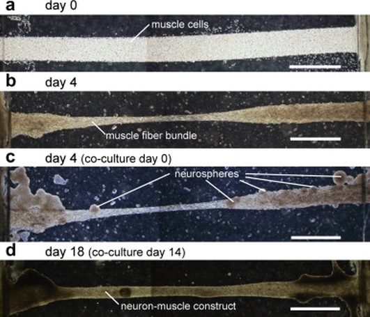
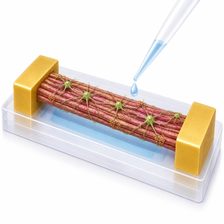
We developed a method for constructing three-dimensional tissues containing neuromuscular junctions by co-culturing motor neurons with engineered skeletal muscle. Neural stem cells were differentiated on free-standing three-dimensional muscle-fiber bundles. The resulting neurites connected to the muscle fibers, where acetylcholine receptors formed at the contact sites. This enabled muscle contraction to be controlled through neuronal signaling rather than direct external electrical stimulation. In the experiments, activation of the neurons with glutamic acid induced large, synchronized contractions of the muscle tissue. Treatment with curare, an inhibitory agent, stopped the movement, demonstrating the formation of functional neuromuscular junctions. This system may contribute to studies of neurodegenerative diseases such as amyotrophic lateral sclerosis, drug development, and biohybrid robots capable of biologically realistic neural control.
Cellular Doll
Molding cell beads for rapid construction of macroscopic 3D tissue architecture
(*equal contribution) Yukiko T.-Matsunaga*, Yuya Morimoto*, Shoji Takeuchi
Advanced Materials, vol. 23, pp. H90-94, 2011
View Research Details
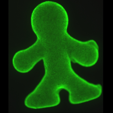
We developed a method for rapidly constructing three-dimensional tissues with arbitrary shapes using cell beads—collagen gel beads coated with cells—as the basic building units. The cell beads were produced using microfluidic technology and poured into a figurine-shaped mold. Cells on the surfaces of neighboring beads adhered to one another, migrated into the interior, and proliferated, forming a mechanically stable tissue within 24 hours. Spaces between the beads allowed nutrients to reach the interior of the construct, helping to prevent central necrosis while maintaining a high cell density even in relatively thick tissues. As a proof of concept, we successfully fabricated a figurine-shaped tissue approximately 5 mm in length. This work introduced an innovative bottom-up approach for constructing complex three-dimensional tissues and organ models.